A considerable number of novice graduate students in research laboratories often inadvertently fall prey to a "gene-centric" cognitive bias. Upon receiving their assigned target gene from their advisor, they eagerly consult all available information regarding the gene—ranging from its sequence and structural configuration to protein-protein interactions, and from its tissue distribution profile to disease associations—meticulously cataloging every detail without discrimination. Yet, after compiling this vast amount of information, they often become disoriented: what aspects of this information are genuinely pertinent to their research? And what precisely should be their next course of action?
A more efficient way of thinking is to adopt a question-centered approach from the very beginning. Your research group focuses on a specific disease or biological process—such as cardiac hypertrophy, liver cancer metastasis, inflammatory response, metabolic disorder, etc.—and the target gene is merely a "handle" or a "probe" to access this scientific question. The goal of the research is not to "thoroughly characterize this gene," but rather to "leverage this gene to answer a biological question related to the disease."
Based on this core concept, this guide constructs a four-stage research framework of "confirming expression differences → verifying functional causality → analyzing downstream mechanisms → refining scientific significance". This framework is driven by specific biological questions rather than being gene-centered, aiming to help researchers establish a clear logical chain: first, clarify whether the gene has changed in the disease scenario, then verify whether this change is the cause driving the disease or a concomitant result, then analyze the underlying molecular regulatory network, and finally refine the theoretical contributions and application values of the research. Following this path, researchers can effectively avoid the common pitfall of "researching a gene for the sake of researching it" and anchor each experimental step on clear scientific questions.
Phase 1: Analysis of the expression profile of the target gene
Phase goal: Clarify whether there are stable and reproducible differences in the expression level of the target gene in the target disease or biological scenario (disease group vs control group).
1.1 Preliminary screening based on public databases
"Before starting wet experiments, it is recommended to prioritize the use of existing public high-throughput data resources:"
1. GEO (Gene Expression Omnibus)
Website:https://www.ncbi.nlm.nih.gov/geo/
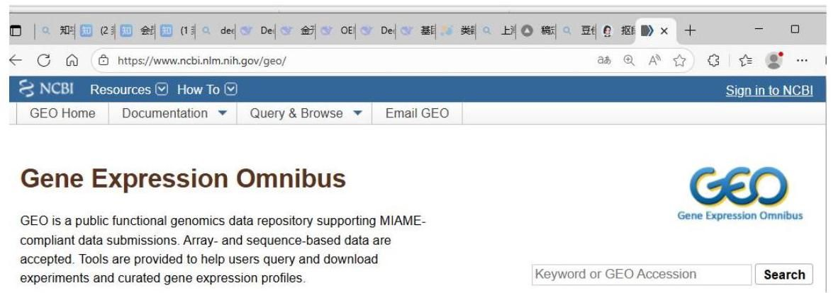
Usage: Retrieve expression profile datasets related to the target disease, screen 2-3 independent cohorts, and examine the differential expression of the target gene between the case group and the control group (box plot, heat map or volcano plot).
Note: An international public database maintained by NCBI that archives high-throughput gene expression and functional genomic data.
2. TCGA (The Cancer Genome Atlas)
Official data portal: https://portal.gdc.cancer.gov
Project homepage: https://www.cancer.gov/ccg/research/genome-sequencing/tcga
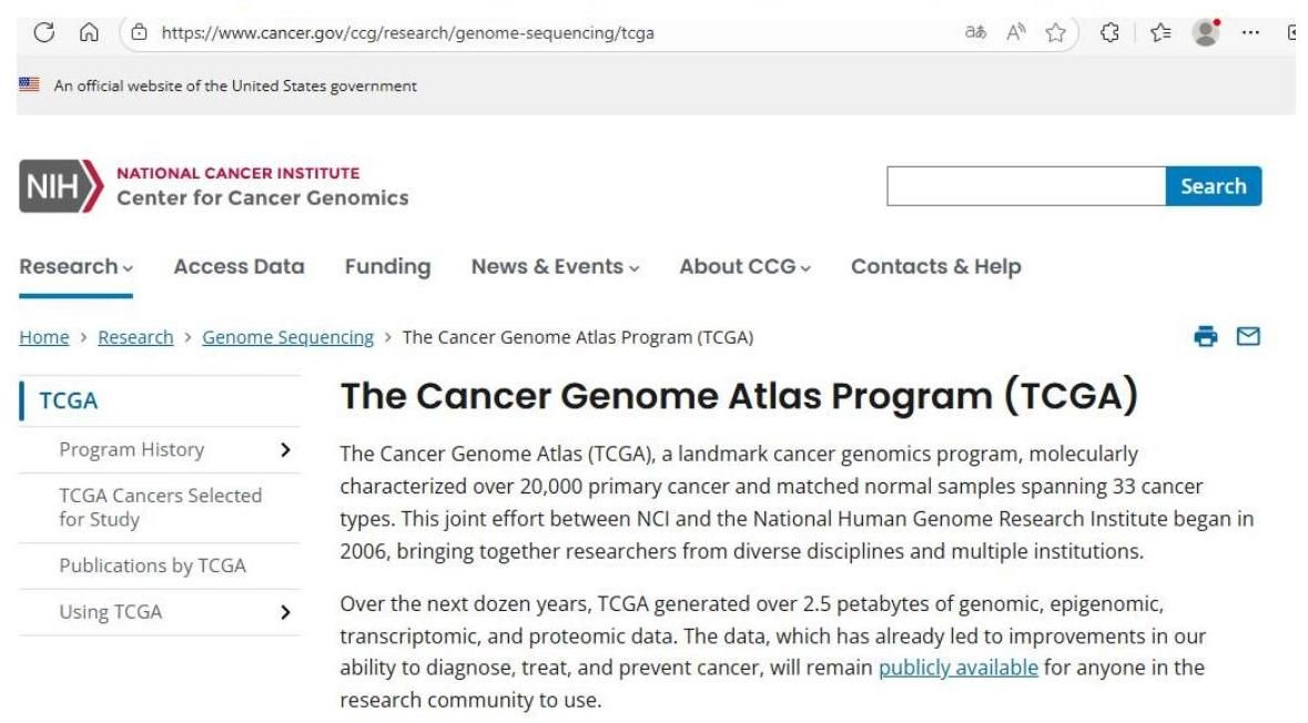
Usage:Applicable to tumor research, for comparing the expression levels of target genes in tumor tissues and paired adjacent cancer tissues. Note: The Cancer Genome Atlas project has performed molecular characterization on more than 20,000 primary cancer samples of 33 cancer types. TCGA data can also be accessed through the UCSC Xena browser (https://xena.ucsc.edu).
3. GTEx (Genotype-Tissue Expression)
Website:https://www.gtexportal.org
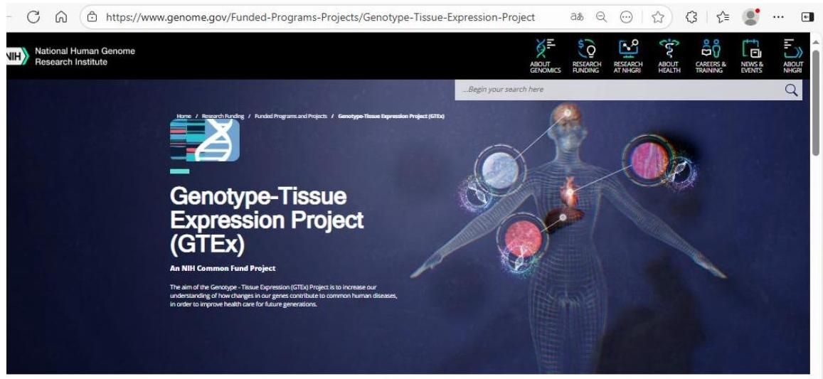
Usage: To obtain the baseline expression distribution of the target gene in various normal human tissues and assist in judging its tissue specificity.
Note: The Genotype-Tissue Expression project has collected approximately 21,000 RNA-seq samples from 54 tissue sites (non-disease sources) for studying the relationship between gene expression and genetic variation.
Through the above analysis, three key pieces of information should be clarified: ① Whether there is reported differential expression of the gene; ② The direction of differential expression (upregulation or downregulation); ③ The magnitude of differential expression and the statistical significance level.
1.2 Experimental Verification Based on Self-Samples
If public data supports the existence of differential expression, or there is a lack of relevant public data, it is necessary to use self-sample resources for verification:
mRNA level:The qPCR technique is used. This method has high sensitivity and low cost and is suitable for preliminary verification. Protein level: Western Blot is used to detect the total protein expression level; immunohistochemistry (IHC) technique is used to detect the in-situ expression and distribution of proteins in tissues; ELISA technique is used to detect the content of secreted proteins in body fluids or culture supernatants.
Subcellular localization: Immunofluorescence (IF) technique combined with confocal microscopy is used to clearly observe the fine localization of the target protein in cells.
Output at this stage: Clearly answer "In the XX disease model, the expression level of target gene A is X times (or reduced to Y% of the control group) that of the normal control state, and the difference is statistically significant ( p<0.05 ), and this difference has been verified in no less than 3 independent biological replicates."
Second Stage: Gain-of-Function and Loss-of-Function Studies of the Target Gene
Stage goal:To determine whether the change in the expression of the target gene is the "cause" driving the phenotypic change or merely an "accompanying phenomenon" in the disease process. This is a key step in establishing the causal relationship between genes and phenotypes.
Core strategy:Exogenously intervene to change the expression level of the target gene and systematically detect the changes in disease-related phenotypes.
|
Scientific question |
Intervention method |
Detection index |
Interpretation ofpositive results |
|
Is overexpression of this gene sufficient to induce disease phenotypes? |
Gain-of-function: Construct an overexpression vector and transduce it into cell or animal models |
Whether the disease-related core phenotypes (proliferation, migration, apoptosis, secretion of inflammatory factors, etc.) deteriorate |
This gene has pathogenic potential |
|
Is knockdown/knockout of this gene sufficient to alleviate disease phenotypes? |
Loss-of-function: Use siRNA, shRNA or CRISPR-Cas9 technology to knockdown/knockout endogenous genes |
Whether the disease-related core phenotypes improve |
This gene is a necessary condition for the occurrence of the disease |
2.1 In vitro cell-level experiments
Model selection:Priority should be given to cell lines with high endogenous expression of the target gene to ensure that the knockdown experiment can observe the dynamic range.
Intervention tools:Use siRNA (transient knockdown) or shRNA (stable knockdown) to construct a knockdown model; use overexpression plasmids or lentiviruses to construct an overexpression model.
Phenotype detection: Select the corresponding detection system according to the disease background -CCK-8 (proliferation), Transwell (migration/invasion), flow cytometry (apoptosis/cell cycle) are commonly used in tumor research; ELISA (inflammatory factor secretion), qPCR (inflammatory gene transcription level) are commonly used in inflammation research; biochemical assays (substrate/product concentration), Seahorse (cellular energy metabolism analysis) are commonly used in metabolism research.
2.2 Animal-level experiments
Intervention methods:Use AAV, lentivirus or adenovirus-mediated in vivo overexpression or knockdown, achieved through local injection (such as intratumoral, stereotactic brain injection) or systemic injection (tail vein, intraperitoneal).
Animal models: Gene knockout (KO) or transgenic (TG) mice that have been constructed can be used; or after establishing a disease model (such as tumor-bearing mice, inflammation models), intervene through virus-mediated gene manipulation.
Observation indicators: Include but are not limited to tumor volume and weight, body weight changes, survival curves, histopathological scores (H&E staining), immunohistochemical detection of marker expression.
2.3 Rescue Experiment
The rescue experiment is the "gold standard" for verifying causal relationships, and the operation process is as follows:
Use siRNA/shRNA to knockdown the expression of the endogenous target gene and observe phenotype improvement;
Under the knockdown background, reintroduce an exogenous target gene (designed to be insensitive to the above siRNA/shRNA, such as introducing silent mutation sites through synonymous mutations);
If exogenous rescue can reverse the phenotype (restore the improved phenotype to the disease state), it strongly proves that the observed phenotype is indeed mediated by the change in the expression of the target gene, rather than caused by off-target effects or other confounding factors.
Output of this stage: Clearly answer "In in vitro and/or in vivo models, upregulating/downregulating target gene A can significantly change the direction and intensity of disease phenotype Y, and the rescue experiment verifies the causal specificity of this effect."
Stage 3: Analysis of downstream molecules and regulatory mechanisms of the target gene
Premise of this stage: It has been confirmed that the change in the expression of target gene A can directly lead to the change in phenotype Y. This stage aims to answer "Through what molecular mechanism does A produce the above phenotypic effect?"
3.1 Prediction of Mechanism Research Strategies Based on Protein Types
The structure and functional characteristics of the protein encoded by the target gene determine its most likely regulatory mode, based on which the corresponding screening strategies can be preferentially selected:
|
Protein type |
Most likely regulatory mode |
Preferred screening technique |
|
Transcription factor (including DNA-binding domain) |
Bind to the promoter region of downstream target genes and regulate their transcription |
RNA-seq + ChIP-qPCR / Dual-luciferase reporter gene |
|
Kinase/Phosphatase |
Catalyze the phosphorylation or dephosphorylation of target proteins and change their activity |
Phosphoproteomics + In vitro kinase assay |
|
Ubiquitin ligase/Deubiquitinase |
Mediate the ubiquitination modification of target proteins and regulate their degradation |
Ubiquitin proteomics + Co-IP verification |
|
Receptor/Channel/Transporter protein |
Bind ligands, transmit signals, and transport substances across membranes |
Co-IP-MS to find interacting proteins + Ligand/substrate identification |
|
Secreted cytokine |
Bind to cell membrane receptors and activate intracellular signal cascades |
Receptor fishing + Screening of signal pathway inhibitor library |
|
Protein with unknown function (no known domain) |
Most likely to function by interacting with proteins with known functions |
Preferred: IP-MS (Immunoprecipitation-Mass spectrometry) |
3.2 High-throughput Screening Strategies
Based on the above judgments, select one or more omics methods for unbiased screening:
Transcriptomics (RNA-seq): Compare the mRNA transcription profiles of the control group and the gene manipulation group (knockdown or overexpression) to identify differentially expressed genes. Further combine GO functional annotation and KEGG pathway enrichment analysis to lock in the significantly enriched biological processes and signaling pathways.
Proteomics (TMT/iTRAQ labeling and quantification): Directly detect the expression changes at the protein level to compensate for the limitation that the transcriptome cannot reflect post-translational regulation.
Interactome proteomics (IP-MS): Use specific antibodies against the target gene for immunoprecipitation to capture protein complexes that directly or indirectly interact with the target protein, and construct an interaction network after mass spectrometry identification.
Alternative Solutions when Funds are Limited:
Database prediction:Use the STRING database to predict protein-protein interaction networks; use the KEGG database to locate the signaling pathways to which the target gene belongs; use the JASPAR database to predict the potential binding sites of transcription factors.
Literature inference:Search in PubMed with the keywords "Gene A + Disease Name + Pathway" to integrate relevant pathway clues in existing reports.
3.3 Causal Verification of Candidate Molecules
From the list of candidate molecules obtained from high-throughput screening, select 2-3 molecules with the highest priority for in-depth verification. The verification needs to answer two progressive questions.
01:Is there a direct physical or regulatory relationship between the target gene A and the candidate molecule B?
|
Hypothetical relationship type |
Verification method |
Criteria for determining positive results |
|
A transcriptional regulation B (A is a transcription factor) |
Dual-luciferase reporter gene assay + ChIP-qPCR |
The activity of the reporter gene increases significantly + ChIP-qPCR shows that the enrichment fold of A in the promoter region of B istimes |
|
A directly binds to B protein |
Endogenous Co-IP (preferred), exogenous Pull-down (in vitro verification), BiFC (live cell verification) |
The antibody of the target protein in endogenous Co-IP can capture B protein simultaneously |
|
A activates a certain signaling pathway (such as NF-κB) |
WB detects the phosphorylation or cleavage level of key proteins in the pathway |
After the operation of A, the activation level of key proteins in the pathway changes in a dose-dependent manner |
|
A affects the synthesis or consumption of a certain metabolite |
Targeted metabolomics + exogenous supplementation experiment |
The metabolite level changes significantly after the operation of A; exogenous supplementation of this metabolite can partially restore the phenotype |
02 :Is B the key mediator for A to regulate phenotype Y? This is the end point of mechanism verification, and a "dual manipulation" functional experimental design should be adopted:
Positive verification (necessity verification): Overexpress A→ phenotype Y enhances → while knocking down B→ . If phenotype Y returns to the baseline level, it proves that B is a necessary mediator for A to mediate the phenotypic effect.
Reverse verification (sufficiency verification): Knock down A→ phenotype Y weakens → while overexpressing B→ . If phenotype Y is restored, it proves that B is a sufficient mediator for A to mediate the phenotypic effect.
Only when positive results are obtained in both of the above verifications can the mechanistic conclusion of "A regulates phenotype Y through B" be drawn.
Stage output: Clearly answer that "the target gene A regulates the downstream molecule B (specific regulation mode: transcriptional activation/protein binding/phosphorylation modification/ubiquitination degradation, etc.), thereby affecting the activity of signaling pathway C, and ultimately leading to changes in phenotype Y . The dual manipulation functional rescue experiment confirms that B is the key mediator molecule for A to regulate Y ."
Phase 4: Evaluation of the biological and translational significance of research findings
Stage goal: Place the stage research findings in the field knowledge system and evaluate their theoretical contributions and potential application values. This stage determines the academic positioning and narrative logic of the research findings.
4.1 Three levels of theoretical contributions
|
Contribution level |
Judgment criteria |
Corresponding statement of scientific significance |
|
Discovery of new mechanism |
There has been no previous report indicating that |
"This study for the first time reveals the regulatory role of the A-B signaling axis in the Y phenotype" |
|
Clinical application potential |
A is differentially expressed in clinical samples and is associated with prognosis; or targeting A/B shows therapeutic effects in animal models |
"Targeting the A/B signaling axis may provide a new strategy for the intervention of this disease" |
|
Revision of theoretical understanding |
It is generally believed in the field that A promotes the disease, and this study demonstrates that A has a protective effect |
"This study presents a conclusion contrary to the original understanding of the function of A in the Y phenotype, suggesting that the pathophysiological role of A needs to be reexamined" |


4.2 Methods for Rapidly Locating the Position of the ResearchDiscovery Field
Search PubMed with the keyword "Gene A + Disease Name" (in English input) and count the number of existing literature:
≥100 papers: A is a "known target" in the field, and "new regulatory mechanisms" or "new phenotypic dimensions" need to be highlighted as innovation points;
- 100 papers: A is a "moderately hot target", and the functional differences and mechanism pathways need to be systematically verified to form a complete evidence chain;
≤10 papers: A is a "novel gene/new association", and the reliability of expression differences and the repeatability of functional importance need to be prioritized and strengthened.
Output at this stage: Be able to clearly state in 1 - 2 sentences "What is the core finding of this study and why this finding is worthy of attention"
Generally speaking, from an unfamiliar gene to a paper, there are actually three logical chains: first, check the basic information, then conduct functional verification, and finally explore the mechanism. Don’t be dazzled by various high - sounding technical terms. The core issues are always two-does this gene have a function? How does it achieve it? If these two things are explained clearly, the research topic is established.