


Esophageal cancer (EC) is the 11th most common malignancy and the 7th leading cause of cancer-related death worldwide, with an overall 5-year survival rate of less than 30%. Esophageal squamous cell carcinoma (ESCC) is its predominant pathological subtype, accounting for more than 85% of all cases. Owing to insidious early symptoms, most patients are diagnosed at an advanced stage. Despite continuous advances in comprehensive therapies including surgery, radiotherapy, chemotherapy and immunotherapy, the risk of recurrence and overall prognosis remain unsatisfactory. Therefore, in-depth exploration of the cellular and molecular mechanisms underlying ESCC progression and identification of novel therapeutic targets have always been core research priorities in this field.
Recently, the research team led by Professor Luowei Wang from the First Affiliated Hospital of Naval Medical University (Changhai Hospital) published findings in Cell Discovery (IF=16.9), entitled FAM50A promotes esophageal squamous cell carcinoma progression via splicing-dependent oncogenic circSDK1- and DUSP22-mediated MAPK/FAK activation. This study systematically uncovered for the first time a novel mechanism by which the splicing factor FAM50A facilitates ESCC progression through the circSDK1-DUSP22 molecular axis, and identified rifabutin as a candidate therapeutic agent.
Notably, the key experiments used to directly verify the interaction between circRNA (circSDK1) and DUSP22 protein in this study — RNA probe synthesis and EMSA (electrophoretic mobility shift assay) — were provided by GeneCreate Biological Engineering Co., Ltd. As a core in vitro assay for validating direct RNA–protein binding, EMSA provided solid experimental evidence for establishing this mechanism.
Key Finding 1: FAM50A is Markedly Upregulated in ESCC and Closely Correlated with Poor Prognosis
By integrating multi-omics data and multiple independent clinical cohorts, the research team systematically screened splicing factors with aberrant expression in ESCC. Combined analysis of transcriptomic data from TCGA-ESCC and GSE53624, as well as proteomic data from two independent cohorts at Changhai Hospital, pinpointed FAM50A among hundreds of splicing factors — a molecule significantly upregulated at both mRNA and protein levels in ESCC.
Clinical correlation analyses revealed that high FAM50A expression was significantly associated with advanced tumor stage and lymph node metastasis. Kaplan–Meier survival analysis demonstrated that patients with high FAM50A expression had markedly shorter overall survival than those with low expression. This finding was consistently validated across TCGA, GSE53624 and the two Changhai Hospital cohorts, strongly indicating that FAM50A acts as a critical oncogenic splicing factor and holds potential as a prognostic biomarker for ESCC.
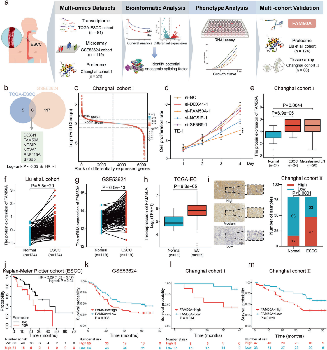
Figure 1. Elevated FAM50A expression correlates with disease progression and poor prognosis in ESCC patients.
Key Finding 2: As a Splicing Factor, FAM50A Regulates Alternative Splicing at the Transcriptome-Wide Level and Promotes circSDK1 Biogenesis
To dissect the molecular mechanism by which FAM50A drives oncogenesis, the team used RIP-seq (RNA immunoprecipitation sequencing) to map the RNA-binding landscape of FAM50A across the whole transcriptome. The analyses showed that FAM50A extensively interacts with core spliceosome components (such as the PRP19 complex and multiple snRNP proteins), and binds numerous mRNAs and non-coding RNAs transcriptome-wide, with binding sites enriched in intronic and promoter regions.
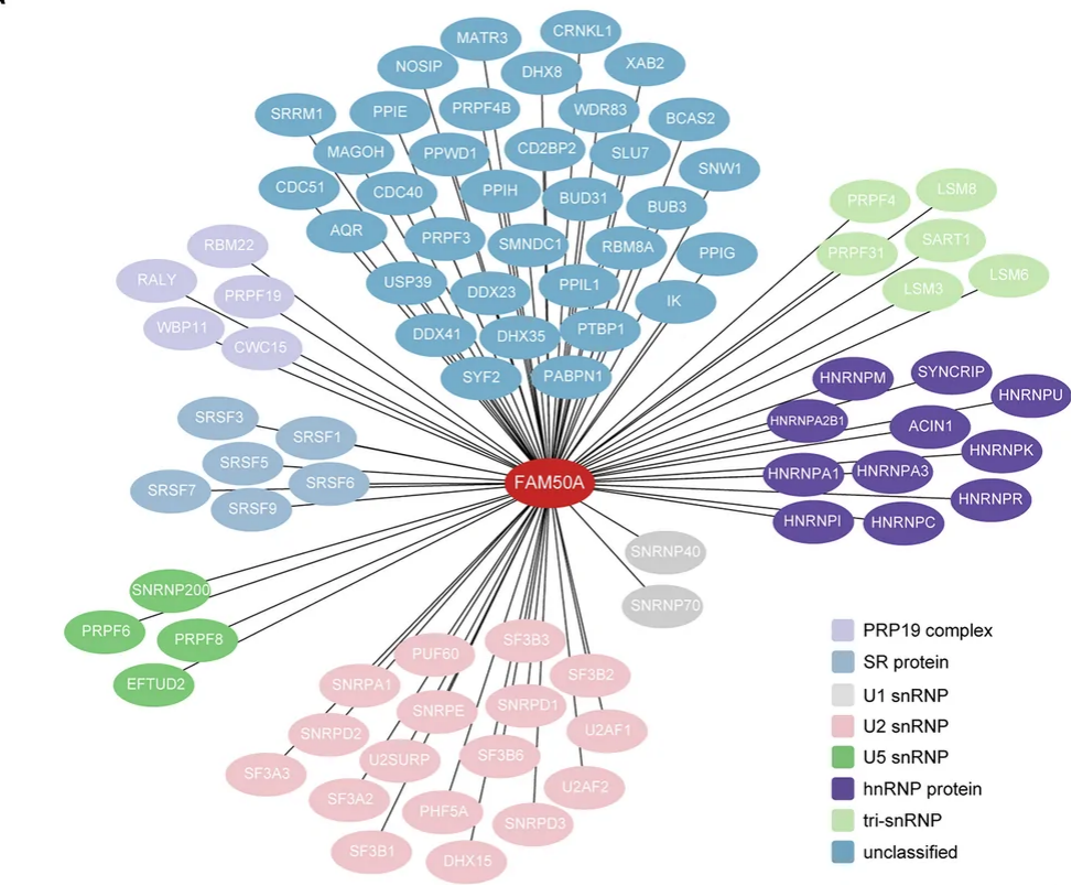
Figure 2. A correlation network between FAM50A and other splicing factors constructed in Cytoscape (nodes are colored by spliceosome components including the PRP19 complex, SR proteins and snRNPs, showing extensive interactions between FAM50A and core spliceosome components).
Further alternative splicing analyses identified substantial alterations in diverse alternative splicing events upon FAM50A knockdown, including exon skipping, alternative 5′ splice sites and alternative 3′ splice sites. Among these differential splicing events, the team focused on a circular RNA, circSDK1. circSDK1 is derived from exons 2–4 of SDK1 pre-mRNA and forms a closed circular structure via back-splicing. RNase R digestion assays and actinomycin D chase experiments confirmed that circSDK1 bears canonical circRNA features: high stability, long half-life, and predominant cytoplasmic localization.
Functional validation experiments showed that circSDK1 expression was drastically reduced after FAM50A knockdown. Silencing circSDK1 significantly suppressed proliferation, migration and invasion of ESCC cells. Reintroduction of circSDK1 into FAM50A-knockdown cells partially rescued the malignant phenotypes. These results demonstrate that circSDK1 serves as a key downstream effector mediating the oncogenic function of FAM50A.
Key Finding 3: circSDK1 Directly Binds DUSP22, Inhibits Its Phosphatase Activity and Sustains MAPK/FAK Activation
How does newly generated circSDK1 exert oncogenic effects? Using RIP-qPCR, RNA pull-down and AlphaFold3 molecular docking analysis, the team narrowed down the candidates and ultimately identified DUSP22 (dual-specificity phosphatase 22) as the downstream interacting protein of circSDK1. DUSP22 is a phosphatase with tumor-suppressive potential that restrains the activity of oncogenic signaling cascades such as MAPK and FAK through dephosphorylation.
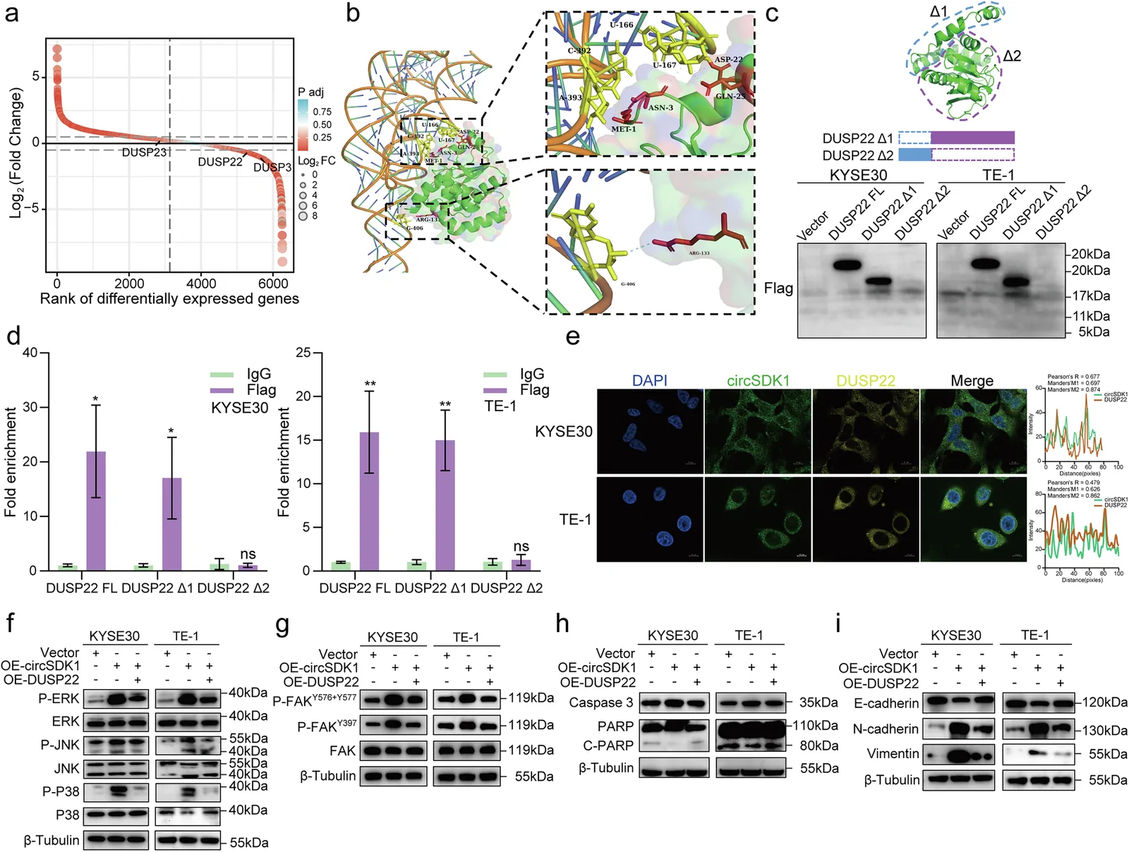
Figure 3. circSDK1 occupies the phosphatase domain of DUSP22 and increases phosphorylation of MAPKs and FAK.
To confirm the direct binding between circSDK1 and DUSP22, the research team further performed EMSA (electrophoretic mobility shift assay). This assay employed purified recombinant DUSP22 protein and in vitro transcribed biotin-labeled circSDK1 RNA probes to directly observe their binding in an in vitro system:
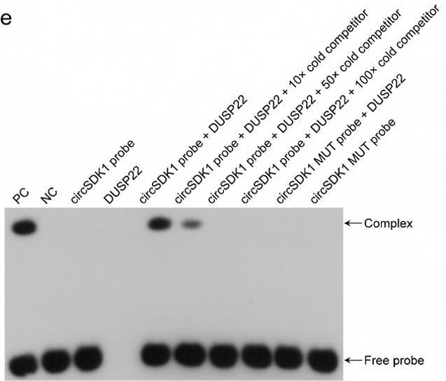
Figure 4. EMSA results: Biotin-labeled probes were used in EMSA to verify the direct interaction between DUSP22 protein and circSDK1.
When biotin-labeled wild-type circSDK1 probe was incubated with DUSP22 protein, a distinct shifted band appeared in electrophoresis, a hallmark of protein–RNA complex formation. Mutant probes failed to form protein–RNA complexes (the shifted band disappeared), indicating that binding depends on specific RNA sequences. Addition of a 10-fold excess of unlabeled wild-type probe (cold competition) partially attenuated the shifted band; a 50-fold or 100-fold excess of cold competitor completely abolished the band, further verifying the specificity of binding.
Collectively, these results confirmed a direct and specific protein–RNA interaction between circSDK1 and DUSP22.
Mechanistically, circSDK1 occupies the phosphatase catalytic domain of DUSP22 and suppresses its dephosphorylation activity. Notably, DUSP22 overexpression reversed the oncogenic effects of circSDK1, suggesting competitive binding between the two. Consequently, downstream MAPK (ERK, JNK, p38) and FAK signaling pathways cannot be properly terminated and remain persistently phosphorylated and activated, which continuously fuels proliferation, migration, invasion and EMT (epithelial–mesenchymal transition) of ESCC cells.
Key Finding 4: TFAP2C Acts Upstream to Regulate FAM50A, and Rifabutin Exhibits Therapeutic Potential
Why is FAM50A highly expressed in ESCC? The team further traced the upstream regulatory axis. Using predictions from databases including JASPAR and TFtarget, combined with dual-luciferase reporter assays and ChIP-qPCR validation, the researchers found that the transcription factor TFAP2C directly binds the promoter region of FAM50A and transcriptionally activates its expression. Analysis of clinical samples also showed that TFAP2C expression positively correlated with FAM50A expression in ESCC tissues, and high TFAP2C expression was likewise associated with poor patient prognosis.
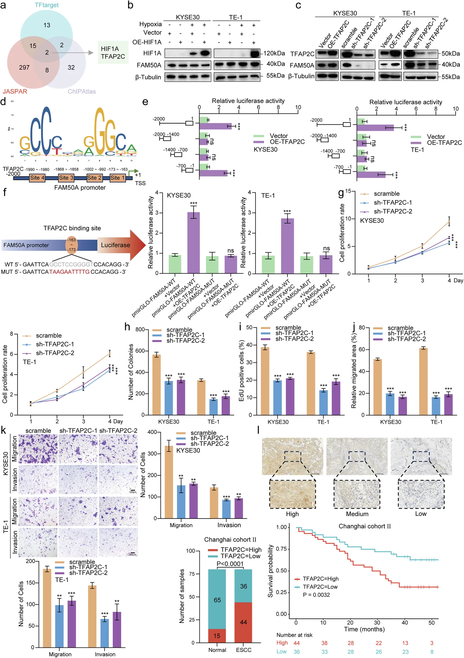
Figure 5. The transcription factor TFAP2C drives transcription of FAM50A, leading to its aberrant upregulation.
Thus, a complete regulatory axis was clearly delineated:
TFAP2C transcriptional activation → FAM50A upregulation → enhanced circSDK1 biogenesis → binding and inhibition of DUSP22 → sustained MAPK/FAK activation → ESCC progression
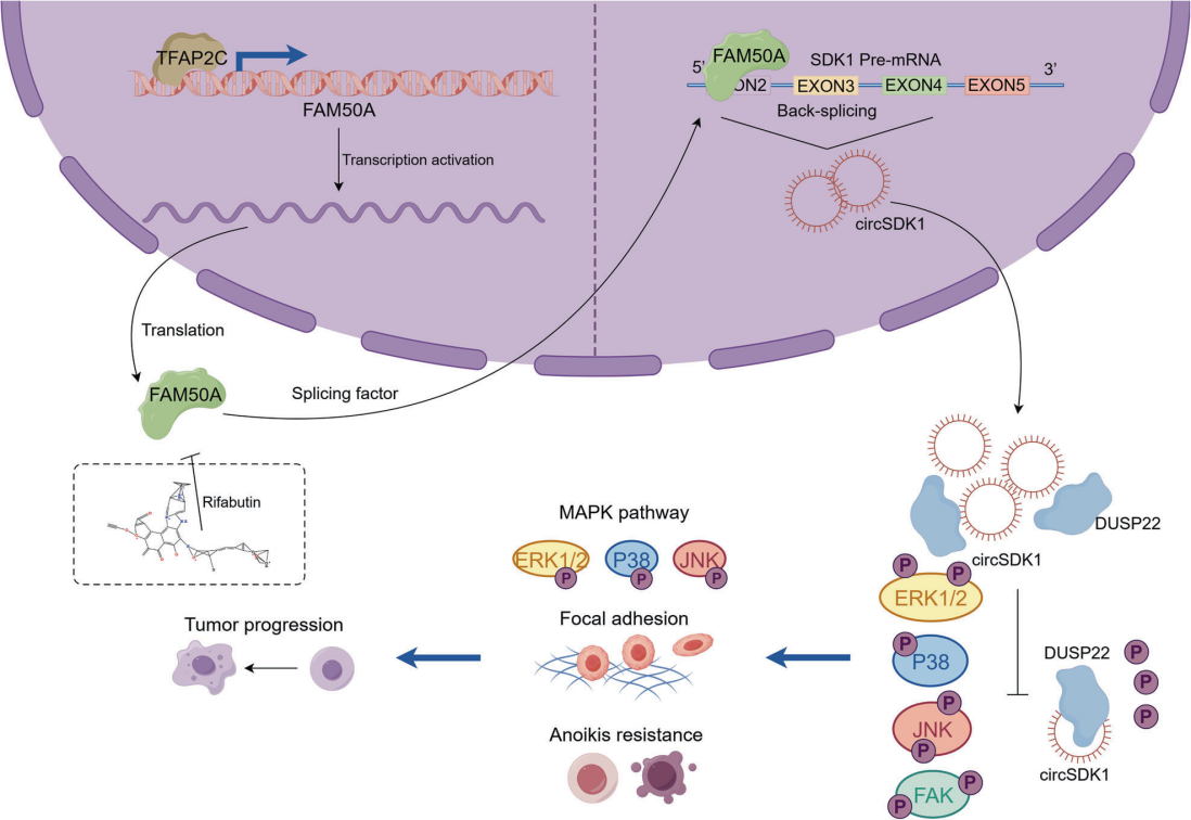
Figure 6. Schematic model: The splicing factor FAM50A promotes back-splicing of SDK1 pre-mRNA to generate circSDK1, which subsequently activates MAPK and FAK signaling pathways.
For translational therapeutics, the team screened an FDA-approved compound library. Using molecular docking and in vitro functional validation, they discovered that rifabutin binds FAM50A protein and inhibits its splicing factor activity. Therapeutic efficacy was validated across multiple model systems: in in vitro cellular assays, rifabutin markedly suppressed ESCC cell proliferation; in cell line-derived xenograft (CDX) models, rifabutin significantly retarded tumor growth; robust anti-tumor effects were also observed in patient-derived organoid (PDO) models and patient-derived xenograft (PDX) models. Moreover, combined treatment with cisplatin suggested synergistic anti-tumor effects.
Significance of This Study
This study systematically revealed for the first time the biological roles and molecular mechanism of the FAM50A/circSDK1/DUSP22 regulatory axis in ESCC, establishing a complete regulatory cascade of “splicing factor–circRNA–phosphatase–signaling pathway”. These findings not only deepen our understanding of ESCC pathogenesis, but also provide novel targets and a candidate drug for the diagnosis and treatment of esophageal squamous cell carcinoma.
About GeneCreate EMSA Service
In this research, the EMSA service provided by GeneCreate supplied experimental data supporting validation of circRNA–protein direct interactions.
EMSA (electrophoretic mobility shift assay) is a classic assay for studying interactions between proteins and nucleic acids (DNA/RNA). By detecting mobility shifts of nucleic acid probes after binding to proteins in gel electrophoresis, direct binding can be intuitively validated. Combined with mutant probes and cold competition assays, the binding specificity can be further confirmed. For studies on interactions between non-coding RNAs (circRNA, lncRNA, etc.) and proteins, EMSA is one of the essential approaches to verify direct binding in vitro.
Beyond EMSA, GeneCreate also offers diverse molecular interaction validation services including ChIP, dual-luciferase reporter assay, RNA pull-down and Co-IP, covering research directions such as transcriptional regulation, epigenetics and non-coding RNA biology.
Feel free to contact us for inquiries if you have relevant experimental requirements.